Collaborative effort by the Suter, Mattoo, and Huang labs provide new insights into filopodia formation
08-20-2019
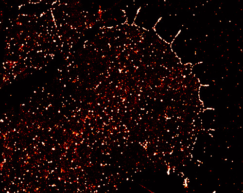
Filopodia are fingerlike structures in neuronal growth cones and many other cells, made up by F-actin bundles. They play important roles for sensing the environment and cell migration. Work in the Suter Lab has recently identified a single tyrosine residue, Y499, in Aplysia cortactin that can be phosphorylated by Src2 and is important for filopodia formation in neuronal growth cones. Phosphorylated cortactin is enriched along the leading edge and filopodia and regulates both filopodia formation and elongation.
These findings have recently been published as the cover article in the July 15 issue of Molecular Biology of the Cell. The research was a collaborative effort by the Suter, Mattoo, and Huang labs at Purdue and was funded by a grant from the National Science Foundation (1146944-IOS) and grants from the Purdue Research Foundation to Daniel M. Suter, grants from the NIH (R35-GM119785) and DARPA (D16AP00093) to Fang Huang, a grant from NIH (R01-GM10092) to Seema Mattoo. Sherlene Brown was supported by a Bird Stair Graduate Student Fellowship from the Department of Biochemistry, Purdue University.
A single tyrosine phosphorylation site in cortactin is important for filopodia formation in neuronal growth cones by Yuan Ren, Yingpei He, Sherlene Brown, Erica Zbornik, Michael J. Mlodzianoski, Donghan Ma, Fang Huang, Seema Mattoo, and Daniel M. Suter.
Abstract
Cortactin is a Src tyrosine phosphorylation substrate that regulates multiple actin-related cellular processes. While frequently studied in nonneuronal cells, the functions of cortactin in neuronal growth cones are not well understood. We recently reported that cortactin mediates the effects of Src tyrosine kinase in regulating actin organization and dynamics in both lamellipodia and filopodia of Aplysia growth cones. Here, we identified a single cortactin tyrosine phosphorylation site (Y499) to be important for the formation of filopodia. Overexpression of a 499F phospho-deficient cortactin mutant decreased filopodia length and density, whereas overexpression of a 499E phospho-mimetic mutant increased filopodia length. Using an antibody against cortactin pY499, we showed that tyrosine-phosphorylated cortactin is enriched along the leading edge.
The leading edge localization of phosphorylated cortactin is Src2-dependent, F-actin–independent, and important for filopodia formation. In vitro kinase assays revealed that Src2 phosphorylates cortactin at Y499, although Y505 is the preferred site in vitro. Finally, we provide evidence that Arp2/3 complex acts downstream of phosphorylated cortactin to regulate density but not length of filopodia. In conclusion, we have characterized a tyrosine phosphorylation site in Aplysia cortactin that plays a major role in the Src/cortactin/Arp2/3 signaling pathway controlling filopodia formation.