New visualization technique has potential to advance structural biology
11-22-2013
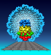
Give several children the exact same set of LEGOs, and each child will create a unique structure. Likewise, a set of biological components can assemble into various structures, and understanding a resulting arrangement is key to understanding its unique function. Dr. Wen Jiang and colleagues developed a new technique to better decipher such structures by focusing sequentially on individual layers.
The new visualization technique, called "focused asymmetric reconstruction" (FAR), refines the previously used "standard asymmetric reconstruction" (SAR). With SAR, the primary component was in focus, but the other parts tended to be blurred. However, FAR varies the focus on each pair of sequential components selectively, blurring the others, allowing for each component to be clearly resolved in relation to its neighbors. Dr. Jiang's team developed the new technique while attempting to visualize a substructure called the portal vertex of a bacteriophage virus (infects bacteria). The portal vertex helps package DNA as it enters the virus' protein shell, a critical step in the life cycle of many viruses. Motivated by the limitations of SAR, the team devised the FAR method to better account for the variety of assemblies created by similar components. Using the new technique, Jiang's team showed that the individual components do not always line up in the same way, as was previously thought, and published their results earlier this year in the Proceedings of the National Academy of Sciences. Other team members included Purdue postdoctoral researchers Fei Guo, and Zheng Liu, graduate students Frank Vago, and Weimin Wu, undergraduate student Yue Ren, as well as collaborators from the University of Texas Health Science Center.
While this is the first clear visualization of the portal under investigation, the FAR technique has other potential applications, expanding the reach of structural biology. Currently, it is most useful for visualizing a stack-like structure, where the primary variations are only along a single axis, much like a combination lock. However, with further development, it could be used to visualize more complex structures.
Guo F, Liu Z, Vago F, Ren Y, Wu W, Wright ET, Serwer P, Jiang W. 2013. Visualization of uncorrelated, tandem symmetry mismatches in the internal genome packaging apparatus of bacteriophage T7. Proceedings of the National Academy of Sciences 110:6811–6816.
http://www.pnas.org/content/110/17/6811
Writer: Karen Wiggins, 49-44749, wiggins@purdue.edu